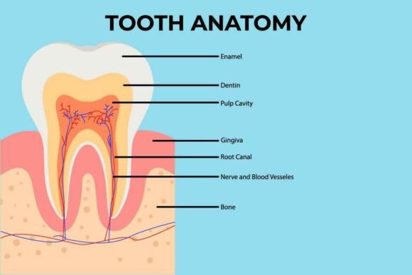
Human Teeth Anatomy Explained
Whether you're designing dental education materials, building a health-focused website, or creating science-based content for schools or clinics, understanding Human Teeth Anatomy isn’t just about memorizing names—it’s about unlocking clarity, credibility, and connection. A well-structured infographic of human teeth anatomy bridges the gap between complex biology and everyday understanding. It transforms dense terminology into visual logic that sticks.
Why This Anatomy Matters Beyond the Dentist’s Chair
Human Teeth Anatomy is more than a clinical reference—it’s a communication tool. Professionals across fields use it to explain oral health risks, illustrate preventive care, support patient consent forms, or even inform product design (like toothbrush ergonomics or orthodontic device interfaces). Educators rely on accurate representations to teach developmental milestones in children; marketers use clean, labeled diagrams to build trust in oral care campaigns; and freelance illustrators integrate anatomical precision to elevate medical storytelling.
Core Structural Layers—What Makes Each Tooth Tick
A single human tooth reveals five functional layers—each with distinct roles:
- Enamel: The hardest substance in the human body—highly mineralized, translucent, and non-regenerative. Its surface texture and thickness affect how light reflects in photography and digital renders.
- Dentin: A porous, yellowish layer beneath enamel. It transmits sensation and supports structural integrity—critical for realistic 3D modeling and cross-sectional illustrations.
- Pulp: The living core housing nerves and blood vessels. Accurate pulp chamber depiction matters for animations showing cavity progression or root canal workflows.
- Cementum: A thin, bone-like covering over the root surface—often overlooked but essential for periodontal ligament attachment visuals.
- Periodontal ligament & alveolar bone: Not part of the tooth itself, but vital context for showing how teeth anchor in the jaw. Omitting them reduces clinical accuracy in surgical or orthodontic infographics.
Real-World Uses You Might Not Expect
Think beyond textbooks. A high-fidelity infographic of human teeth anatomy serves tangible needs across sectors:
- Educators embed static or animated versions into LMS platforms—students retain up to 40% more when layered diagrams replace bullet-point lists.
- Dental startups use annotated tooth structure visuals in investor decks to demonstrate product alignment with biological realities (e.g., biomimetic filling materials).
- Content creators repurpose modular elements—like isolated enamel cross-sections or erupting molar sequences—as social media carousels, boosting engagement without oversimplifying.
- Freelance designers license scalable vector versions for white-label client work—ensuring consistency across brochures, apps, and explainer videos.
Design Decisions That Impact Usability
Not all Human Teeth Anatomy infographics deliver equal value. Look for these markers of quality:
- Proportional accuracy: Molars shouldn’t appear oversized next to incisors unless explicitly illustrating comparative function.
- Consistent labeling hierarchy: Primary terms (e.g., “enamel”) in bold, secondary descriptors (e.g., “hydroxyapatite crystals”) in smaller type—no visual noise.
- Color logic: Use clinically aligned palettes—not arbitrary brights. Enamel = off-white/pearl, dentin = warm ivory, pulp = soft coral. This builds subconscious recognition and avoids misinterpretation.
- Adaptability: Vector-based files (SVG, EPS) scale cleanly for print posters or retina-display web banners—raster-only PNGs limit flexibility.
Where Missteps Happen—and How to Avoid Them
Even experienced teams misjudge nuance. Common pitfalls include:
- Showing deciduous (baby) teeth alongside permanent teeth without clear age context—confusing caregivers reading pediatric oral hygiene guides.
- Using mirrored left/right views without side labels—problematic for surgical prep or orthodontic planning where laterality is critical.
- Oversimplifying root morphology: A maxillary first molar often has three roots; a mandibular premolar usually has one—but variations exist. Good infographics note “common” vs. “variable” features.
- Ignoring occlusion lines or wear patterns—missed opportunities to show real-world function, like how enamel thins at biting edges over time.
Practical Tips for Selecting or Commissioning Visuals
If you’re sourcing or creating an infographic of human teeth anatomy, ask yourself:
- Does it reflect current ADA or FDI World Dental Federation nomenclature? (e.g., “first molar” not “6-year molar”)
- Are eruption timelines included—or at least referenced—for developmental context?
- Can key structures be isolated (via layers or transparency) for custom presentations?
- Is there version control? Updated editions should reflect new research—like recent findings on dentin fluid dynamics or enamel remineralization pathways.
Ultimately, Human Teeth Anatomy isn’t static data—it’s living information. When translated thoughtfully into visual form, it supports better decisions: a parent choosing fluoride toothpaste, a designer optimizing a teledentistry UI, a blogger explaining gum recession, or a clinician walking through treatment options. The strongest infographics don’t shout “science”—they invite curiosity, clarify complexity, and earn trust by honoring both accuracy and accessibility. That balance is what makes Human Teeth Anatomy a quietly powerful asset—not just for dentists, but for anyone translating health knowledge into action.